Osteogenesis, ossification, remodeling and growth of bone
(Redirected from Ossification)
Phylogenetically dual bone development:
- primary (covering bone)
- secondary (replacement bones)
Primary Bones[edit | edit source]
- an ossification from ligament = desmogenous ossification
Secondary Bones[edit | edit source]
- at first-cartilaginous
- chondrogenic ossification
Chondrogenic (deeper) and desmogenous (superficial, covering) were often combined to form larger combined units.
Both methods of bone formation were preserved in human embryonic development:
- ossification desmogenous = endesmal (in ligament)
- chondrogenic ossification - division into two groups according to the place of origin in the cartilage:
- perichondrial − surface ossification from the perichondrium
- enchondral − ossification inside the cartilage
Ossification process[edit | edit source]
- osteoblasts − cells from the mesenchyme, produce non-calcified precursors of the ground substance → turn into osteoid by polymerization
- osteoblasts become stuck in this mass
- bone beams − structures that are created by osteoblasts, further increase by apposition
- osteocytes − typical bone cells, it arises from immobile osteoblasts (which are stuck in osteoid)
- osteoclasts - break down bone
- broken bone is replaced by new bone → bone remodeling
- Bone breakdown occurs in two ways. On the basis of the genetic program, the bone is given the desired shape and on the basis of its loading, the internal architecture is formed in the spongiosa.
Desmogenous ossification[edit | edit source]
- new formation of bone beams directly in the ligament → endesmally
- first fibrous bone, which is then rebuilt into cancellous bone
- bones of the cranial vault, facial part of the skull
Chondrogenic ossification[edit | edit source]
- it replaces the original cartilaginous bone model, which is destroyed by this bone formation
Ossification of long bones[edit | edit source]
Perichondral ossification[edit | edit source]
Begins in the perichondrium and occurs only in the diaphysis. The deposition of new bone under the periosteum. Growth in width.
Bone remodeling: continues throughout life
o Osteoclasts remove old bone in tunnel-like cavities
o Capillaries and osteoprogenitor cells get into the tunnels from the endosteum and periosteum
o Osteoblasts develop on the walls of the tunnels and secrete osteoid
o The osteoblasts differentiate to osteocyte and the lamellar bone is formed
Bone repair – in case of fracture
o blood vessels are torn and release blood that clots and produces a large fracture hematoma
o the hematoma is removed by macrophages and replaced by a soft fibrocartilage-like tissue of callus (unorganized network of woven bone) rich with collagen fibers
*if the periosteum is broken it will reestablish continuity over the fibroblast tissue
o the soft callus is invaded by new blood vessels and osteoblasts, resulting with the fibrocartilage being replaced by woven bone o the woven bone is later replaced with lamellar bone
*in children the fracture heals faster due to the ‘green stick fracture’ where the bone bends and cracks (the periosteum doesn’t break) and not broken into fragments
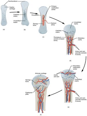
Endochondral ossification[edit | edit source]
Pre-existing hyaline cartilage is being replaces by osteoblasts into bone. Longitudinal growth.
- In the 1st trimester, a collar of bone tissue is formed around the diaphysis of the cartilage model, by osteoblasts existing the perichondrium.
- The collar prevents oxygen and nutrients supply to the cartilage tissue 🡪 enhanced degeneration: the chondrocytes swell and compress their matrix, leading to the calcification and death of the chondrocytes
- The periosteal bud penetrates the periosteum (former perichondrium) and introduces blood vessels and osteoprogenitor cells to the primary ossification center
- The osteoblasts produce woven bone while the osteoclasts degrade cartilage matrix. The bone continues to elongate. Chondrocyte produce cartilage matrix 🡪 osteoclast degrades it 🡪 osteoblasts deposit bone matrix
- The osteoclast degrades most of the spongy bone center creating the medullary cavity, where we can later find the bone marrow
- Around birth secondary ossification centers appear in the epiphysis and develop similarly
- Growth (epiphyseal) plates − between the epiphyses and the diaphysis, they are maintained throughout the bone growth (disappears around the age of 18)
- only in them the bone grows in length - the cartilage creates new material
- one of them (proximal or distal) is more involved in bone growth than the other (e.g. proximal is more active in the humerus and lower leg bones, distal is more active in the forearm and femur)
- at the same time, the bone beams break down in the diaphysis → cavitas medullaris, a medullary cavity is created.
- growth in thickness − by apposition from the periosteum and partly also from the endosteum
- apposition must be supplemented by bone resorption (remodeling)
Areas of ossification (from epiphysis to diaphysis)[edit | edit source]
● Zone of reserve cartilage: a hyaline cartilage that is not affected by the bone formation
● Zone of growth: chondrocytes proliferate and form columns
● Zone of hypertrophic cartilage: the chondrocytes enlarge, compressing the matrix
● Zone of maturation: where calcification occurs
● Zone of ossification: calcium reaches from the diaphysis, further calcifying the chondrocytes. They are then degraded by osteoclasts and replaced with bone by osteoblasts
Ossification of short bones[edit | edit source]
- Enchondrally - in the middle of the cartilaginous base
- spread of ossification to the bone surface (occurs throughout the growth period)
- formation of perichondral mantle lamella (at the end of the growth period) → formation of superficial compact bone
Ossa pneumatica[edit | edit source]
- the mucous membrane of the nasal cavity is inserted into some skull bones after birth
- the mucous membrane of the middle ear cavity is inserted into part of the temporal bone
- the spongiosa of the bone recedes in front of the mucous membrane, the cavity enlarges
- with the original cavity (nasal, middle ear) from which the mucous membrane has been removed, the cavity in the pneumatic bone remains connected only by a narrow passage
Links[edit | edit source]
Related Articles[edit | edit source]
Reference[edit | edit source]
- ČIHÁK, Radomír. Anatomie. 2.. edition. Grada, 2001. ISBN 80-7169-970-5.


